- Case report
- Open access
- Published:
Bilateral chronic cavitary pulmonary aspergillomas in an adult patient with recurrent tuberculosis: a case report and literature review
Journal of Medical Case Reports volume 18, Article number: 491 (2024)
Abstract
Background
Aspergillomas are globular growths of Aspergillus fumigatus, a benign aspergillosis of the lungs. It usually affects patients who are immunocompromised and have anatomically defective lung structures. The majority of aspergilloma cases are asymptomatic, despite the fact that 10% of cases spontaneously resolve. Most patients do not have any symptoms from their lesions. Direct serological or microbiological evidence of an Aspergillus species along with radiologic evidence is required for the diagnosis of an aspergilloma.
Case
We describe a 35-year-old adult Oromo male patient who had been experiencing night sweats, an intermittent productive cough with sparse whitish sputum, loss of appetite, and easy fatigability for 3 months. At 5 years prior, he received treatment for pulmonary tuberculosis that was smear-positive and was subsequently certified healed. Objectively, he was tachypneic and had intercostal, subcostal, and supraclavicular retractions with symmetric chest movement. A high-resolution computed tomography scan revealed bilateral apical cavitary lesions with core soft tissue attenuating spherical masses and an air crescentic sign suggestive of aspergillomas, which were confirmed by sputum light microscopic examination. The patient was managed with antibiotics and antifungals.
Conclusion
Aspergilloma is a symptom of chronic pulmonary aspergillosis, a category of lung disorders caused by a persistent Aspergillus infection. Primary aspergillomas are uncommon and frequently occur in people with compromised immune systems. A prolonged cough, fever, chest pain, and hemoptysis are all symptoms of pulmonary aspergillomas. The majority of the time, pulmonary aspergillosis is difficult to identify. Despite high mortality and morbidity rates, surgery is still the most effective treatment for pulmonary aspergilloma.
Introduction
Aspergillomas are globular masses of Aspergillus fumigatus, a noninvasive form of pulmonary aspergillosis [1]. The genus Aspergillus has approximately 40 species that can cause infection [2], with A. fumigatus, A. flavus, and A. terreus being the most common causes of invasive aspergillosis in immunocompromised people. These species produce conidia in concentrations ranging from 1 to 100 conidia per m3 [3]. Patients with anatomically defective lungs and preexisting cavities develop aspergilloma. Primary aspergillomas are uncommon and typically develop in people with compromised immune systems, such as those with neutropenia, long-term glucocorticoid users, or acquired immunodeficiency syndrome (AIDS), as a result of Aspergillus species invading the bronchi and causing cavitation [4]. When immunocompetent patients are exposed to Aspergillus spores in dry or extremely dusty surroundings, hay barns, or compost sites and have prior lung diseases, such as tuberculosis (TB), sarcoidosis, lung abscess, bronchogenic cysts, or lung tumor, secondary aspergilloma develops [5,6,7]. The most frequent preexisting lung disease is tuberculosis, and there can be a delay of from less than 1 year up to 30 years between TB and aspergilloma diagnoses [6]. Denning et al. reported the involvement of Aspergillus infection in pulmonary TB and asthmatic patients. It is projected that at least 372,385 people globally develop chronic pulmonary aspergillosis after being treated for pulmonary tuberculosis [8]. The natural history of aspergilloma is still mostly unknown [9]. Although 10% of aspergilloma cases spontaneously resolve, the majority of cases remain asymptomatic [9, 10]. Even with treatment, pulmonary aspergilloma still has a high rate of morbidity and mortality. If left untreated, damage may eventually engulf an entire lobe or lung [11, 12]. Chest imaging may show some distinctive patterns that help with this disease’s diagnosis, although pathological tissue examination is frequently required for a certain diagnosis [13]. Hemoptysis, cough, chest pain, and fever are some of the nonspecific symptoms [14]. Severe or persistent hemoptysis caused by the intrapulmonary lesion is fatal [15], because antifungal medications only partially eliminate the fungal ball that has settled down in the pulmonary cavities [16]. In addition to symptom relief, surgical removal of the circumscribed pulmonary lesion increases the possibility of aspergilloma lasting remission [17, 18]. Below we discuss an adult patient with tuberculosis recurrence who was additionally diagnosed with bilateral chronic cavitary aspergillomas. This case entails a unique presentation, as the co-occurrence of active pulmonary tuberculosis and pulmonary aspergillomas is quite rare and requires a high index of suspicion and the timely initiation of antituberculosis and antifungal agents. Furthermore, the index case brings a learning point and consolidates the existing clinical knowledge of infectious diseases, the importance of imaging and options for management.
Case presentation
This is a 35-year-old Oromo male patient who presented with intermittent productive cough with scanty whitish sputum, night sweating, unquantified but significant weight loss, loss of appetite, and easy fatigability of 3 months duration. The above symptoms worsened over the past 3 weeks, with associated symptoms, such as high-grade fever, exertional dyspnea, and an increased amount of sputum, which is sometimes blood tingled. He had a history of treatment for smear-positive pulmonary tuberculosis 5 years ago and was declared cured after treatment. Otherwise, he had no history of smoking cigarettes, orthopnea, paroxysmal nocturnal dyspnea or body swelling, or chronic medical illnesses, such as diabetes and hypertension. On physical examination, he was acutely sick looking (in cardiorespiratory distress). His vital signs were blood pressure of 107/70 mmHg, pulse rate of 124 beats per minute, respiratory rate of 32 breaths per minute, axillary temperature of 38.7 °C, and peripheral oxygen saturation of 85% with room air. On respiratory system examination he was tachypneic, had intercostal, subcostal, and supraclavicular retractions with symmetrical chest movement. There was relative dullness over posterior lower one third of left lung. On auscultation, there were fine crackles over upper two thirds of both lungs posteriorly and absent air entry over posterior lower one third of left lung.
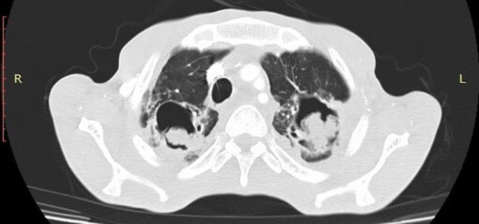
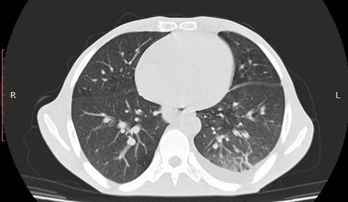
He was investigated with hematological tests, chemistry and imaging. Accordingly, his white blood cell counts = 9000/uL, hemoglobin = 15gm/dl, neutrophil = 81%, random blood sugar = 134gm/dl, creatinine = 0.64, urea = 143, alkaline phosphatase = 100 IU/L, aspartate aminotransferase (AST) = 19 IU/L, alanine aminotransferas (ALT) = 26 IU/L, human immunodeficiency virus (HIV) test = non-reactive, erythrocyte sedimentation rate (ESR) = 74 mm/hour, and sputum geneXpert rifampicin resistance (RIF) assay showed that mycobacterium tuberculosis was detected. However, rifampicin resistance was not detected. Electrocardiography showed normal sinus rhythm. Posteroanterior chest x-ray was taken and it showed bilateral upper lung old fibrotic changes, bilateral upper lung zones pulmonary nodules, and left lower lung zone consolidation with pleural effusion (Fig. 1). High resolution chest computed tomography (CT) scan was taken and revealed bilateral apical lungs cavitary lesions with central area soft tissue attenuating rounded masses surrounded by an air crescent sign suggestive of aspergillomas (Fig. 2). Left lung consolidation with bilateral lower lobe ground glass opacities and left pleural collection secondary to bronchopneumonia with parapneumonic effusion (Fig. 3). A light microscopic examination of a sputum sample revealed broad, colorless, septated, and branching hyphae. Mycological culture and serological tests were not done owing to unavailability of these tests at our institution.
Later on, with assessment of bilateral chronic cavitary pulmonary aspergillomas plus post tuberculosis lung fibrosis plus severe community acquired pneumonia with para pneumonic effusion plus pulmonary tuberculosis (relapse), the patient was admitted to male medical ward and started management. He was put on intranasal oxygen at 5 L/minute, ceftriaxone at 1 gm intravenously twice a day for 7 days, ciprofloxacin at 400 mg intravenously twice a day for 10 days, azithromycin at 500 mg orally daily for 5 days, anti-TB 2RHZE/4RH regimen (four tablets daily), pyridoxine at 25 mg orally daily, and itraconazole at 400 mg orally daily. After 2 weeks of commencement of the above management, he was discharged with improvement. Objectively, the vital signs were all in normal range. On chest examination, there was decreased air entry over the posterior lower one-third of the left lung field. He was discharged with anti-TB, itraconazole at 200 mg orally twice a day for 6 months, pyridoxine at 25 mg orally daily for 1 month, and had a monthly appointment. He was counseled on the necessary precautions that should be taken to prevent transmissions of tuberculosis to his family members and the community. He was reevaluated 1 month after discharge, and his respiratory condition significantly improved though he complained intermittent shortness of breath, cough, and easy fatigability. The repeated chest x-ray showed remission of consolidation and pleural effusion, although there was no change in the cavitary lesion. CT scan was not repeated because the patient was not comfortable paying for the imaging for the second time. He was advised to take his medication strictly and to continue his follow-up regularly.
Discussion
Aspergilloma is a symptom of chronic pulmonary aspergillosis, a group of illnesses brought on by persistent lung infection with Aspergillus [19]. A single (simple) pulmonary aspergilloma is a fungal ball that grows in a single lung cavity. Chronic cavitary pulmonary aspergillosis, formerly known as complex aspergilloma, is characterized by many cavities that may or may not contain an aspergilloma, as well as pulmonary and systemic symptoms and elevated inflammatory markers, over at least 3 months of monitoring [11, 20]. Many Aspergillus species are common saprophytes in nature. Aspergillus fumigatus is mostly responsible for pulmonary illness. In wealthy nations, such as the USA, the prevalence of chronic pulmonary aspergillosis is lower, with less than 1 case per 100,000 people [21]. John Hughes Bennett first described cavitary Aspergillus disease in 1842, explaining the condition as having soft tuberculous materials in several cavities of various sizes in the lung. Aspergilloma was first fully characterized and given the term “mega mycetoma” by Deve in 1938 [22]. Later, Hinson and colleagues characterized aspergilloma as a saprophytic infection of preexisting lung cavities, which is how it is currently understood [23]. Aspergillus spores primarily enter the body through the respiratory system, while they can also live as commensals in the external auditory canal. These filamentous organisms then multiply quickly and cause serious problems in a sick lung or when there is a systemic immune deficit [24]. Aspergillus manufactures the proteins gliotoxin, fumagillin, and helvoic acid to evade the host elimination system. In endothelial and epithelial cells, the protein generated can restrict cilia motility and promote conidia internalization. Conidia can connect with endothelial cells, infiltrate tissue hyphae, and engage with leukocytes in the respiratory system [25, 26]. The adaptive immune response in humans that is responsible for conidia clearance in immunocompetent hosts, as well as where conidia colonize in immunocompromised hosts, is poorly known [27].
There are a number of risk factors that can potentially lead to the development of pulmonary aspergilloma. These are pulmonary tuberculosis, cystic fibrosis, chronic bronchiectasis, pneumoconiosis, post infarct pulmonary cavity, post radiation pulmonary cavity, sarcoidosis, bronchial cysts and bullae, chronic lung abscess, lung malignancy, ankylosing spondylitis, malnutrition, chronic obstructive pulmonary disease (COPD), chronic liver disease, post-transplant, stem cell transplant, chemotherapy, neutropenia, prolonged corticosteroid use, HIV, and primary immunodeficiency syndromes [24, 28, 29]. From all of these, TB is the one that is the most frequently linked with pulmonary aspergilloma [30]. There is also new evidence of chronic cavitary pulmonary aspergillomas following coronavirus disease 2019 (COVID-19) pneumonia. A recent report stated that a 64-year-old man was treated for COVID-19 with tocilizumab and dexamethasone. Then, 3 months later, he revisited the health facility, complaining of hemoptysis, a productive cough, and weight loss [31]. Similarly, pulmonary aspergillomas has been reported in a patient receiving teriflunomide for multiple sclerosis [32]. It was believed that the development of aspergillomas in people with healthy immune systems required a structural alteration to the nest that caused the airflow to become stagnant, allowing Aspergillus to colonize [33]. Our case was treated for sputum positive pulmonary tuberculosis 5 years ago. Even though he was declared cured, currently his sputum gene expert result is positive for tuberculosis. Previous exposure to tuberculosis might have caused lung fibrosis. Post tuberculosis fibrotic lung is the main risk factor leading to the development of aspergillomas. Even though the vast majority of papers on pulmonary aspergilloma mentioned the occurrence of aspergilloma following previous pulmonary tuberculosis, there are no works of literature stating concomitant occurrence of tuberculosis recurrence with pulmonary aspergillomas.
The majority of patients with aspergilloma probably do not experience any symptoms from their lesions. When present, the symptoms might vary and are frequently challenging to identify because of other underlying pulmonary disease processes [34]. Chronic cough, fever, chest pain, hemoptysis, and certain symptoms related to chronic wasting disorders are among the clinical manifestations of pulmonary aspergilloma [35]. Hemoptysis is the most typical symptom of aspergilloma [36]. The fungus’s endotoxins, local invasion of the blood vessels bordering the cavity, or mechanical stimulation of the exposed vasculature inside the cavity by the rolling fungus ball can all cause bleeding, which typically comes from bronchial blood vessels [37]. The intercostal arteries themselves could also be the source of bleeding. Intercostal artery erosion may result from the mycotic process spreading and causing parenchymal damage at the lung’s periphery to invade the nearby chest wall [38]. Large artery bleeding is unlikely to stop on its own and may be fatal. Between 2 and 14% of people die from aspergilloma-related hemoptysis [15, 39]. It is impossible to determine which patients would develop life-threatening hemoptysis on the basis of the magnitude, complexity, presence of warning mild hemoptysis, or kind of underlying disease [15]. Our patient, manifested with intermittent productive cough for 3 months associated with loss of appetite and weight. In addition, he had night sweating, high grade fever, and exertional dyspnea. Although he seldom saw blood-tinged sputum, he did not report to have frank hemoptysis.
Most of the time, it can be difficult to diagnose pulmonary aspergillosis. To diagnose an aspergilloma, direct serological or microbiological evidence of an Aspergillus species should be combined with radiologic evidence. Serodiagnosis approaches employing galactomannan and d-glucan, on the other hand, have limited sensitivity and specificity [40]. An Aspergillus-specific immunoglobulin G (IgG) antibody assay outperformed traditional precipitant antibody assays in terms of sensitivity and repeatability. It is now the most reliable approach for detecting chronic cavitary pulmonary aspergillomas (CPA) produced by Aspergillus fumigatus, but there is no evidence that it is efficient in identifying CPA caused by non-fumigatus Aspergillus [41]. Mycological culture is also commonly used approach for diagnosing CPA, however it has significant drawbacks. According to reports, the culture positivity rates of Aspergillus species from respiratory specimens can be as low as 11.8% [40, 42, 43]. Despite the fact that clinical signs including weight loss, a productive cough, hemoptysis, shortness of breath, chest tightness, and fever can be helpful, these symptoms are general and may be linked to the underlying pulmonary disease [44]. In CT scans of the chest, aspergillomas may be seen in the pulmonary, pleural, or ecstatic bronchus. It is the most defining imaging aspect of chronic pulmonary aspergillosis [45].The characteristic radiological sign of aspergilloma is a movable, intracavitary mass that typically develops in the upper lobes; the mass is almost pathognomonic for the condition [46]. As the patient is moved, it may display the Monod sign, a rounded mass that typically travels inside the cavity, and provide the traditional “air crescent sign” [47]. Radiographically, in contrast to simple aspergillomas, which may have thin walls, normal adjacent lung parenchyma, and no pleural involvement, chronic cavitary pulmonary aspergillomas may be more aggressive, causing more significant destruction of the lung parenchyma, poorly defined consolidation regions, and multiple cavities containing fungus balls, debris, and fluid [9, 45, 47]. Patients with simple aspergilloma are frequently asymptomatic; however, those with chronic aspergilloma frequently exhibit more severe symptoms, such as hemoptysis, bronchorrhea, chest discomfort, inadequate nutrition status, and decreased respiratory function [48, 49]. We did a microscopic test of sputum sample, which showed a septated and branching hyphae that in turn indicates the presence of fungal infection. Since we do not have serological tests for fungal infections in our set up, the tests were not performed. We did PA chest x-ray and high-resolution chest CT scan. The imaging findings were consistent with chronic cavitary aspergillomas. The pathognomonic radiological signs, such as Monod sign and air crescent signs, were clearly visible in this particular case.
Despite high rates of death and morbidity, surgery is still the best treatment for pulmonary aspergilloma. This claim is supported by the recent retrospective study in Algeria involving 69 patients who were operated for pulmonary aspergillomas with 75% mortality [50]. Surgically, the fungal ball, the underlying cavity, and the diseased parenchyma around it can all be removed simultaneously. The purpose of surgery is to prevent a potentially fatal hemoptysis as well as the invasive clinical forms of pulmonary fibrosis and renal amyloidosis that are brought on by persistent inflammation [51]. It is necessary to determine whether the patient is operable before deciding on the surgical method and to plan for various postoperative consequences by evaluating respiratory function and comorbidities [52]. Poor respiratory reserve, multiple aspergillomas or bilateral aspergillomas, and patient preference are among conditions that make a patient contraindicated for surgery [44]. There are various options available for inoperable patients that need to be treated medically. Amphotericin B is claimed to have a cure rate of 10% when administered systemically, which is comparable to the rate of spontaneous resolution [16]. Likewise, this choice is undesirable owing to the dangers of amphotericin B, specifically nephrotoxicity. Nevertheless, intralesional infiltration of amphotericin B has brought a dramatic clinical improvement in patient with chronic cavitary aspergillomas according to a recent report from Portugal, where the procedure involved multidisciplinary health care workers owing to recurrent hemoptysis and refusal of the patient for blood transfusion [53]. Owing to its relatively low cost, oral route of administration, significant lung penetration, and potent activity against aspergillus fumigatus, itraconazole is the azole that has undergone the most testing to treat aspergillomas [54]. Similar to the treatment of invasive pulmonary aspergillosis, the Infectious Disease Society of America (IDSA) advises itraconazole at a dose of 200 mg orally every 12 hours, with the total period of treatment dependent on the person’s clinical and radiographic response [55]. Voriconazole is another oral agent and is used for treating resistant strains of Aspergillus fumigatus. For pulmonary aspergilloma, the IDSA advises voriconazole at doses of 6 mg/kg intravenously every 12 hours for the first day and 4 mg/kg intravenously every 12 hours after that. Oral therapy can be 200–300 mg every 12 hours [55]. Azoles can not be used as the primary treatment for aspergilloma owing to a number of drawbacks. Itraconazole’s overall effectiveness is less than 70%, while voriconazole’s efficacy has not yet been determined. Moreover, it typically takes more than 6 months of treatment to completely eradicate the infection, and incidences of aspergilloma recurrence after stopping the antifungal medication have been reported [56]. We treated our case medically with itraconazole owing to the fact that the fungal balls are bilateral and absence of an experienced cardiothoracic surgeon in our institution. The patient was simultaneously treated for pulmonary tuberculosis and aspergillomas. He tolerated the burden of medications and showed significant functional improvement over subsequent follow-up.
Conclusion
Aspergilloma is a sign of chronic pulmonary aspergillosis, a group of diseases caused by a persistent Aspergillus infection in the lungs. Primary aspergillomas are rare and often appear in patients with immune systems that are already impaired. Clinical signs of pulmonary aspergilloma include a persistent cough, fever, chest discomfort, and hemoptysis. Most of the time, pulmonary aspergilloma is difficult to diagnose requiring high index of suspicion and conducting proper laboratory tests and imaging. There are two options of management of chronic cavitary pulmonary aspergillomas; medical and surgical methods. Antifungal drugs, such as azoles, have good cure rate compared with other drugs with less side effects and the choice should be made on an individual basis. Despite high mortality and morbidity rates, surgery remains the best treatment for simple pulmonary aspergilloma.
Availability of data and materials
All data and materials for this case report are available from the corresponding author upon reasonable request.
Abbreviations
- AIDS:
-
Acute immunodeficiency syndrome
- CT:
-
Computed tomography
- COPD:
-
Chronic obstructive pulmonary disease
- COVID-19:
-
Corona virus disease 2019
- CPA:
-
Chronic pulmonary aspergillomas
- HIV:
-
Human Immunodeficiency virus
- IDSA:
-
Infectious disease society of America
- PA:
-
Postero-anterior
- RH:
-
Rifampicin, isoniazid
- RHZE:
-
Rifampicin, isoniazid, pyrazinamide, ethambutol
- TB:
-
Tuberculosis
References
Kambouris ME, et al. Beyond the microbiome: germ-ganism? An integrative idea for microbial existence, organization, growth, pathogenicity, and therapeutics. OMICS J Integr Biol. 2022;26(4):204–17.
Verweij P, Brandt M. Aspergillus, fusarium, and other opportunistic moniliaceous fungi. Manual Clin Microbiol. 2006;2:1802–38.
Barnes PD, Marr KA. Aspergillosis: spectrum of disease, diagnosis, and treatment. Infect Dis Clin. 2006;20(3):545–61.
Martinez R, et al. Primary aspergilloma and subacute invasive aspergillosis in two AIDS patients. Rev Inst Med Trop Sao Paulo. 2009;51:49–52.
Lachanas E, et al. An unusual pulmonary cavitating lesion. Respiration. 2005;72(6):657.
Chen J-C, et al. Surgical treatment for pulmonary aspergilloma: a 28 year experience. Thorax. 1997;52(9):810–3.
Jameson JL et al. Harrison’s principles of internal medicine. (No Title), 2018.
Denning DW, Pleuvry A, Cole DC. Global burden of chronic pulmonary aspergillosis as a sequel to pulmonary tuberculosis. Bull World Health Organ. 2011;89(12):864–72.
Moodley L, Pillay J, Dheda K. Aspergilloma and the surgeon. J Thorac Dis. 2014;6(3):202.
Kant S, Verma S. Fungal ball presenting as Haemoptysis. Internet J Pulm Med. 2007;10:e1–4.
Denning DW, et al. Chronic cavitary and fibrosing pulmonary and pleural aspergillosis: case series, proposed nomenclature change, and review. Clin Infect Dis. 2003;37(Supplement_3):S265–80.
Saraceno JL, et al. Chronic necrotizing pulmonary aspergillosis*: approach to management. Chest. 1997;112(2):541–8.
Zmeili OS, Soubani A. Pulmonary aspergillosis: a clinical update. J Assoc Phys. 2007;100(6):317–34.
Pohl C, et al. Pulmonary aspergilloma: a treatment challenge in sub-Saharan Africa. PLoS Negl Trop Dis. 2013;7(10): e2352.
Jewkes J, et al. Pulmonary aspergilloma: analysis of prognosis in relation to haemoptysis and survey of treatment. Thorax. 1983;38(8):572–8.
Hammerman KJ, Sarosi GA, Tosh FE. Amphotericin B in the treatment of saprophytic forms of pulmonary aspergillosis. Am Rev Respir Dis. 1974;109(1):57–62.
Belcher J, Plummer N. Surgery in broncho-pulmonary aspergillosis. Br J Dis Chest. 1960. https://doi.org/10.1016/S0007-0971(60)80067-8.
Battaglini JW, et al. Surgical management of symptomatic pulmonary aspergilloma. Ann Thorac Surg. 1985;39(6):512–6.
Lee SH, et al. Clinical manifestations and treatment outcomes of pulmonary aspergilloma. Korean J Intern Med. 2004;19(1):38.
Denning DW, et al. Chronic pulmonary aspergillosis: rationale and clinical guidelines for diagnosis and management. Eur Respir J. 2016;47(1):45–68.
Addrizzo-Harris DJ, et al. Pulmonary aspergilloma and AIDS: a comparison of HIV-infected and HIV-negative individuals. Chest. 1997;111(3):612–8.
McCarthy D, Pepys J. Allergic broncho-pulmonary aspergillosis: clinical immunology:(1) Clinical features. Clin Exp Allergy. 1971;1(3):261–86.
Hinson KF, Moon AJ, Plummer NS. Broncho-pulmonary aspergillosis; a review and a report of eight new cases. Thorax. 1952;7(4):317–33.
Chakraborty RK, Baradhi KM. Aspergilloma, in StatPearls. 2022, StatPearls Publishing.
Hohl TM, Feldmesser M. Aspergillus fumigatus: principles of pathogenesis and host defense. Eukaryot Cell. 2007;6(11):1953–63.
Soewondo W, et al. Co-existing active pulmonary tuberculosis with aspergilloma in a diabetic patient: a rare case report. Radiol Case Rep. 2022;17(4):1136–42.
Shankar J. An overview of toxins in Aspergillus associated with pathogenesis. Int J Life Sci Biotechnol Pharm Res. 2013;2(2):16–31.
Hsiao C-W, et al. Complex pulmonary aspergilloma: a case report. J Med Sci. 2001;21(6):301–4.
Smith N, Denning D. Underlying conditions in chronic pulmonary aspergillosis including simple aspergilloma. Eur Respir J. 2011;37(4):865–72.
Kawamura S, et al. Clinical evaluation of 61 patients with pulmonary aspergilloma. Intern Med. 2000;39(3):209–12.
Adetiloye AO, et al. A 64-year-old man hospitalized for COVID-19 pneumonia and treated with tocilizumab who developed chronic cavitary pulmonary aspergillosis. Am J Case Rep. 2023;24: e938359.
London F, Stanciu-Pop C, Mulquin N. Chronic cavitary pulmonary aspergillosis in a teriflunomide-treated multiple sclerosis patient. Clin Neurol Neurosurg. 2024;236: 108125.
Ma JE, et al. Endobronchial aspergilloma: report of 10 cases and literature review. Yonsei Med J. 2011;52(5):787–92.
Flye MW, Sealy WC. Pulmonary Aspergilloma: a report of its occurrence in 2 patients with cyanotic heart disease. Ann Thorac Surg. 1975;20(2):196–203.
Fujiuchi S, et al. Clinical analyses of Aspergillus infections in patients with underlying pulmonary disease. Nihon Kokyuki Gakkai Zasshi J Jpn Respir Soc. 2004;42(10):865–70.
Faulkner SL, et al. Hemoptysis and pulmonary aspergilloma: operative versus nonoperative treatment. Ann Thorac Surg. 1978;25(5):389–92.
Solit RW, et al. The surgical implications of intracavitary mycetomas (fungus balls). J Thorac Cardiovasc Surg. 1971;62(3):411–22.
Young VK, et al. Operation for cavitating invasive pulmonary aspergillosis in immunocompromised patients. Ann Thorac Surg. 1992;53(4):621–4.
Garvey J, et al. The surgical treatment of pulmonary aspergillomas. J Thorac Cardiovasc Surg. 1977;74(4):542–7.
Kitasato Y, et al. Comparison of Aspergillus galactomannan antigen testing with a new cut-off index and Aspergillus precipitating antibody testing for the diagnosis of chronic pulmonary aspergillosis. Respirology. 2009;14(5):701–8.
Takazono T, Izumikawa K. Recent advances in diagnosing chronic pulmonary aspergillosis. Front Microbiol. 2018;9:1810.
Kohno S, et al. Intravenous micafungin versus voriconazole for chronic pulmonary aspergillosis: a multicenter trial in Japan. J Infect. 2010;61(5):410–8.
Shin B, et al. Serum galactomannan antigen test for the diagnosis of chronic pulmonary aspergillosis. J Infect. 2014;68(5):494–9.
Lang M, et al. Non-surgical treatment options for pulmonary aspergilloma. Respir Med. 2020;164: 105903.
Roberts CM, Citron KM, Strickland B. Intrathoracic aspergilloma: role of CT in diagnosis and treatment. Radiology. 1987;165(1):123–8.
Fishman A. Pulmonary diseases and disorders. Tuberous Sclerosis Lymphangiomyomatosis. 1988;2:965.
Sharma S, et al. ‘Monod’ and ‘air crescent’ sign in aspergilloma. Case Rep. 2013;2013:2013200936.
Gossot D, et al. Full thoracoscopic approach for surgical management of invasive pulmonary aspergillosis. Ann Thorac Surg. 2002;73(1):240–4.
Lejay A, et al. Surgery for aspergilloma: time trend towards improved results? Interact Cardiovasc Thorac Surg. 2011;13(4):392–5.
Ghebouli K, et al. Pulmonary aspergilloma surgery analysis and results of a series of 69 cases. EAS J Med Surg. 2024;6(02):36–40. https://doi.org/10.36349/easjms.2024.v06i02.005.
Liebler JM, Markin CJ. Fiberoptic bronchoscopy for diagnosis and treatment. Crit Care Clin. 2000;16(1):83–100.
Britton NG, Stagg M. Tests of pulmonary function before thoracic surgery. Anaesth Intensive Care Med. 2017;18(12):598–601.
Pinto M, et al. Endobronchial amphotericin B to treat hemoptysis in an inoperable patient with aspergillosis. Med Mycol Case Rep. 2024;43: 100627.
Gupta PR, Jain S, Kewlani JP. A comparative study of itraconazole in various dose schedules in the treatment of pulmonary aspergilloma in treated patients of pulmonary tuberculosis. Lung India. 2015;32(4):342.
Patterson TF, et al. Practice guidelines for the diagnosis and management of aspergillosis: 2016 update by the Infectious Diseases Society of America. Clin Infect Dis. 2016;63(4):e1–60.
Campbell J, et al. Treatment of pulmonary aspergilloma with itraconazole. Thorax. 1991;46(11):839–41.
Acknowledgements
We would like to thank the patient for allowing us to write this case report.
Funding
We received no financial support for this case report.
Author information
Authors and Affiliations
Contributions
TMT conceived and designed the project and carried out data curation, manuscript writing, editing, analysis, and patient follow-up; OS, SDA, EMT, BS, and MTJ carried out supervision and validation. All authors read and approved the final manuscript.
Corresponding author
Ethics declarations
Ethics approval and consent to participate
There are no ethical concerns about this case report.
Consent for publication
Written informed consent was obtained from the patient for publication of this case report and any accompanying images. A copy of the written consent is available for review by the Editor-in-Chief of this journal.
Competing interests
We declare that there are no competing interests.
Additional information
Publisher’s Note
Springer Nature remains neutral with regard to jurisdictional claims in published maps and institutional affiliations.
Rights and permissions
Open Access This article is licensed under a Creative Commons Attribution-NonCommercial-NoDerivatives 4.0 International License, which permits any non-commercial use, sharing, distribution and reproduction in any medium or format, as long as you give appropriate credit to the original author(s) and the source, provide a link to the Creative Commons licence, and indicate if you modified the licensed material. You do not have permission under this licence to share adapted material derived from this article or parts of it. The images or other third party material in this article are included in the article’s Creative Commons licence, unless indicated otherwise in a credit line to the material. If material is not included in the article’s Creative Commons licence and your intended use is not permitted by statutory regulation or exceeds the permitted use, you will need to obtain permission directly from the copyright holder. To view a copy of this licence, visit http://creativecommons.org/licenses/by-nc-nd/4.0/.
About this article
Cite this article
Tadesse, T.M., Shegene, O., Abebe, S.D. et al. Bilateral chronic cavitary pulmonary aspergillomas in an adult patient with recurrent tuberculosis: a case report and literature review. J Med Case Reports 18, 491 (2024). https://doi.org/10.1186/s13256-024-04801-y
Received:
Accepted:
Published:
DOI: https://doi.org/10.1186/s13256-024-04801-y


