- Case report
- Open access
- Published:
Familial adenomatous polyposis: a case report
Journal of Medical Case Reports volume 18, Article number: 415 (2024)
Abstract
Background
Familial adenomatous polyposis is characterized by the presence of multiple colorectal adenomatous polyps and caused by germline mutations in the tumor suppressor gene and adenomatous polyposis coli, located on chromosome 5q21–q22. Familial adenomatous polyposis occurs in approximately 1/10,000 to 1/30,000 live births, and accounts for less than 1% of all colorectal cancers in the USA. It affects both sexes equally and has a worldwide distribution. The incidence of colon cancer in low- and middle-income countries is rising. In addition to the increasing incidence, lack of early detection and impeded access to optimal multidisciplinary treatment may worsen survival outcomes. Developing quality diagnostic services in the proper health context is crucial for early diagnosis and successful therapy of patients with colorectal cancer, and applying a resource-sensitive approach to prioritize essential treatments on the basis of effectiveness and cost-effectiveness is key to overcoming barriers in low- and middle-income countries. We report a case of familial adenomatous polyposis presenting as adenocarcinoma with multiple colorectal adenomatous polyps. The diagnosis of familial adenomatous polyposis was made by the presence of numerous colorectal adenomatous polyps and family history of colonic adenocarcinoma. Due to its rarity, we decided to report it.
Case presentation
A 22-year-old Ethiopian female patient presented to Addis Ababa University College of Health science, Addis Ababa, Ethiopia with rectal bleeding. Abdominopelvic computed tomography scan was done and showed distal rectal asymmetric anterior wall thickening in keeping with rectal tumor. Colonoscopy was done and she was diagnosed to have familial adenomatous polyposis with severe dysplasia. In the meantime, colonoscopy guided biopsy was taken and the diagnosis of adenocarcinoma with familial adenomatous polyposis was rendered. For this, total proctocolectomy was carried out. On laparotomy there was also incidental finding of left ovarian deposition for which left salpingo-oophorectomy was done, and 4 weeks after surgical resection, the patient was started on oxaliplatin, leucovorin, fluorouracil chemotherapy regimen.
Conclusion
In the clinical evaluation of a patient with rectal bleeding, familial adenomatous polyposis must be considered as a differential diagnosis in subjects having family history of colonic adenocarcinoma for early diagnostic workup, management, family genetic counseling, and testing.
Background
Familial adenomatous polyposis (FAP) is an autosomal dominant disease caused by mutations in the APC gene. Classic FAP is characterized by the presence of 100 or more adenomatous colorectal polyps. When fully developed, patients can have up to thousands of colorectal adenomas and a 100% risk of colorectal cancer. Screening for tumors associated with FAP should be performed in individuals with a pathogenic APC mutation. Screening for FAP-associated cancers should also be performed in individuals at risk for FAP who have either not undergone genetic evaluation or have indeterminate genetic test results. Screening for colorectal cancer and other FAP-associated cancers in these patients must be individualized on the basis of their personal and family history of adenomas and cancer. Individuals at risk for FAP include first-degree relatives of those with FAP and individuals with > 10 cumulative colorectal adenomas or colorectal adenomas in combination with extracolonic features associated with FAP (for example, duodenal/ampullary adenomas, desmoid tumors, papillary thyroid cancer, congenital hypertrophy of the retinal pigment epithelium, epidermal cysts, or osteomas) [1]. Polyps occur in the upper gastrointestinal tract in 83–100% of patients with FAP [2, 3].
Considering the increasing number of locally advanced and advanced cases of colorectal cancer (CRC) in low- and middle-income countries (LMICs), there is an urgent need to implement screening strategies for early disease detection. Screening programs are aimed at early detection, recognizing early signs and symptoms of the presence of the disease, and treating patients with curative intent. Screening programs were reported to be more effective in slow-growing cancers with a natural history of multistage progression, such as the adenoma-carcinoma sequence in CRC. Delays in the diagnosis of CRC are multifactorial. There are social, cultural, and structural reasons such as poverty, the misbelief of the incurability of any tumor, the fear of stigma (especially in women), and structural barriers related to proper health facility accessibility due to long distances or unaffordable cancer services not covered by national health schemes of insurance [4, 5]. There is a lack of standard diagnostic facilities in Ethiopia, generating a cancer delay in Ethiopia, which causes increased mortality due to locally advanced presentation. There are a limited number of gastroenterologists and colonoscopy units in Ethiopia, primarily located in urban areas, leaving the rural population needing access to such diagnostic facilities. Due to the shortage of workforce and endoscopic facilities, training programs need to be developed.
Primary assessment of rectal bleeding includes: careful attention to history, presence or absence of perianal symptoms, age of patient (in view of likely differential diagnosis with each age group), family history of colorectal malignancy, and red flag symptoms such as weight loss, symptoms suggestive of anemia, and change in bowel habits [6].
Examination of the abdomen to exclude abdominal mass and digital rectal examination to examine for fissure and exclude rectal cancer may be useful. Fecal calprotectin is a useful screening tool in younger, lower risk patients with suspected inflammatory bowel disease. A positive fecal calprotectin result has a high positive predictive value for finding inflammatory bowel disease at colonoscopy. Proctoscopy is useful for primary care clinicians as a screening tool in patients with rectal bleeding; however, it should not be used as a substitute for flexible sigmoidoscopy to rule out serious pathology. Secondary assessment of rectal bleeding includes: localization of the site and determination of the cause of bleeding to allow for treatment to be appropriately focused. The cause and site of massive lower gastrointestinal hemorrhage should be determined following the early use of colonoscopy and use of computed tomography, computed tomography angiography, or digital subtraction angiography. Flexible sigmoidoscopy is the investigation of choice for patients < 45 years old with persistent rectal bleeding who have received treatment for hemorrhoids and still have persistent bleeding. If there is a family history of colorectal malignancy, colonoscopy may be a better investigation for rectal bleeding as these patients have a higher risk of right colon cancers. Patients > 45 years old with persistent rectal bleeding should be offered either colonoscopy or flexible sigmoidoscopy [6].
Herein, we report a case of FAP presenting as rectal bleeding that was clinically considered as ulcerative colitis. As a result, we are reporting this case due to its rarity and to emphasize the importance of considering FAP in the differential diagnosis of rectal bleeding for early diagnostic workup, management, family genetic counseling, and testing.
Case presentation
A 22-year-old Ethiopian female patient presented to Addis Ababa University College of Health science, Addis Ababa, Ethiopia, with a complaint of on and off type of rectal bleeding of 2-year duration with recent worsening 6 months prior to her current admission. She had visited a nearby health center on multiple occasions for this complaint and took unspecified medications, but experienced no improvement. There was no history of fever, cough, weight loss, night sweating, or loss of appetite. She was having mild abdominal distension, vomiting, abdominal pain, bloody diarrhea, and constipation starting 1 week prior to her current admission. She had family history (first degree relatives) of colon cancer but no history of diabetes or hypertension. Her past medical history was not significant. She had no previous history of admission to hospital. She had no history of any form of surgical procedures. She was not married and lived with her parents. On physical examination, there was mild abdominal tenderness, pale conjunctiva, and nonicteric sclera. On the basis of the above findings, a provisional clinical impression of ulcerative colitis was entertained.
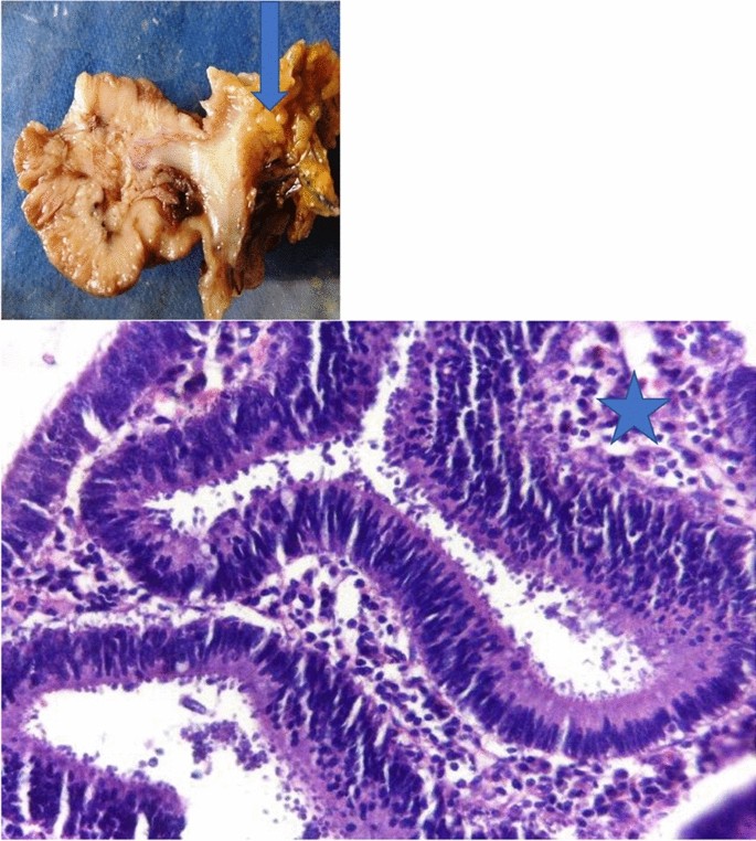
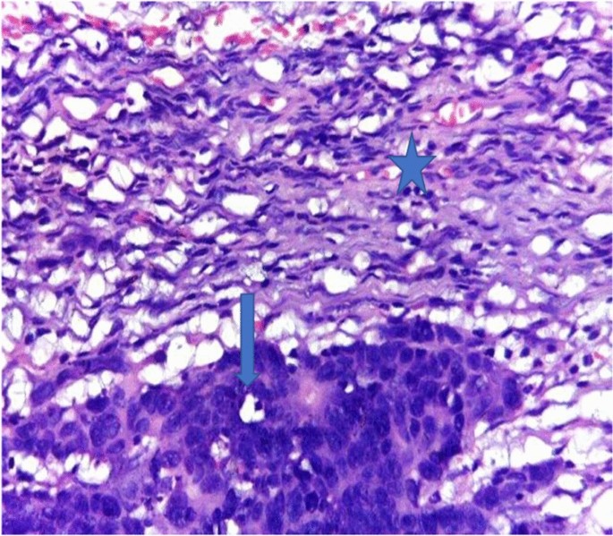
Liver was not palpable below costal margin. There was no splenomegaly. Other clinical findings were within normal limits. Laboratory investigations carried out on the same day of her presentation, including complete blood count (CBC), erythrocyte sedimentation rate (ESR), and chest and abdominal X-ray, were noncontributory. On CBC, total white blood cell (WBC) count was 4000 µL with 50% granulocytes, 45% lymphocytes, 1% eosinophils, and 4% monocytes. Platelet count was 300,000 µL. Hemoglobin was 11.5 g/dL with mean corpuscular volume (MCV) of 75 fL. ESR was 14 mm/hour. Renal function test revealed blood urea nitrogen (BUN) of 12 mg/dL, and serum creatinine level was 0.68 mg/dL. On liver function test, total bilirubin was 0.6 mg/dL, serum albumin was 4.2 g/dL, and serum aspartate transaminase (AST/SGOT) and serum alanine transaminase (ALT/SGPT) were 28 IU/L and 30 IU/L, respectively. Serum electrolytes were in the normal range. Repeated carcinoembryonic antigen (CEA) level was 25 ng/mL. Urinalysis was also done and it was normal. Sputum was negative for acid-fast bacilli. Serum was negative for human immunodeficiency virus (HIV) antibody. Abdominopelvic CT scan was done and showed distal rectal asymmetric anterior wall thickening in keeping with rectal tumor. Uterus, liver, spleen, bilateral ovaries, and kidneys were free of tumor. Colonoscopy was done and reveals numerous colorectal adenomatous polyps; in the meantime, colonoscopy guided biopsy was carried out and the patient was diagnosed with adenocarcinoma. Due to limited number of surgeons and long waiting list of patients, she underwent total proctocolectomy 1 week after her initial presentation. On laparotomy there was also incidental finding of left ovarian deposition, for which left salpingo-oophorectomy was carried out. The specimen was sent to pathology department for gross and histopathologic examination. Gross cut surface examination of proctocolectomy specimen showed numerous adenomatous polyps involving rectum and the entire colon, while there was gray white solid infiltrating mass on left salpingo-oophorectomy specimen (Figs. 1, 2, 3, 4).
Microscopic examinations from both adenomatous polyps and infiltrating gray white solid mass of left salpingo-oophorectomy specimen showed proliferations of highly pleomorphic round-to-oval to polygonal cells having hyperchromatic nuclei and frequent mitotic activities forming variable sized glands admixed with desmoplastic stroma (Figs. 5, 6, 7). Out of 20 lymph nodes sampled, 7 were involved by tumor. On the basis of above findings, the case was diagnosed as adenocarcinoma with lymph node and left ovary metastasis plus FAP. The patient had good postoperative condition, and 4 weeks after surgical resection, she was started on FOLFOX (oxaliplatin 85 mg/m2 intravenous, leucovorin 400 mg/m2 intravenous, fluorouracil 400 mg/m2 intravenous bolus then 2400 mg/m2 intravenous administered over 46 hours) chemotherapy regimen every 2 weeks for 12 rounds. Since then, she was followed up with regular serum CEA, CBC, organ function tests and abdominopelvic CT scan. The patient was having a smooth course with no significant adverse effects encountered. Currently the patient has completed her chemotherapy regimen and is doing well.
Discussion
There is an urgent need for screening strategies for the early detection of colorectal cancer in LMICs, with delays in diagnosis due to social, cultural, and structural barriers. Cost considerations play a role in the success of screening programs, and primary prevention strategies such as education and healthy living are essential. Screening policies for CRC require the engagement of medical leaders, advocacy groups, education, and national cancer control plans. Investment opportunities in the healthcare system need to be identified to maximize benefits. There is a lack of standard diagnostic facilities, imaging techniques, and pathology reporting consensus in most low-income countries, causing a delay in cancer diagnosis and increased mortality due to locally advanced presentation; and while molecular biomarkers are increasingly utilized in management, their implementation in LMICs should prioritize those that are clinically useful, validated, and cost-effective, and building partnerships with HICs for developing research precision biomarkers laboratory and cost-effective strategies should be part of future planning for LMICs. To improve CRC care in LMICs, there is a need to promote clinical research and include clinical data from LMICs in international literature, expand clinical trials, and establish research collaborations between HICs and LMICs. Improving the infrastructure for diagnosis, surgery, and medical and radiation oncology, as well as ensuring access to essential chemotherapy drugs and palliative cancer care is necessary to provide optimal treatment to patients with CRC in LMICs, and should be incorporated into national health policies with adequate funding. Primary prevention strategies are essential in educating the general population, including a healthy diet and living, physical activity, avoiding smoking and alcohol use, and encouraging them to participate in CRC screening. Implementing these primary prevention strategies in LMICs at a population level may control risk factors of noncommunicable disease. We may also have to focus on populations with a high risk of developing colorectal tumors. Well-defined inherited syndromes such as Lynch’s syndrome or familial adenomatous polyposis can occur in 2–5% of CRCs.
Education needs to be improved, and it is crucial to provide access to genetic testing to identify high-risk populations for screening and provide primary preventive surgery or personalized endoscopic plans [7]. Although there are few private sector clinical laboratories in Ethiopia that help clinicians in molecular diagnosis of cancer, the majority of patients cannot afford the high cost of molecular studies. The same was true for our patient.
Practical management of lower gastrointestinal (LGI) bleeding depends on the severity of the hemorrhage and the availability of diagnostic and therapeutic methods at the admitting facility. Endoscopic and radiological techniques have improved to the point that the site of bleeding can be localized in the majority of cases. In addition, episodes of LGI bleeding are less serious than upper gastrointestinal bleeding, with an 80% rate of spontaneous cessation of bleeding and a lower mortality of 2–4% [8] versus 6–13% [9]. It is therefore appropriate to stabilize the patient hemodynamically for transfer to a larger center if expertise in noninvasive diagnostic and therapeutic interventions is not available locally. Similarly, if the bleeding has stopped spontaneously and all investigations are noncontributory, supportive medical management can be continued with repetition of examinations if bleeding recurs. While there is no clear consensus for management as there is for upper gastrointestinal (UGI) bleeding, the following course of management can be proposed on the basis of an overview of all the diagnostic and therapeutic modalities and in accordance with the recommendations of the French Society of Digestive Endoscopy (SFED), American Gastroenterological Association (AGA), and American Society for Gastrointestinal Endoscopy (ASGE) [10,11,12]. Proposed management of acute LGI and chronic LGI bleeding with no hemodynamic instability is shown in Figs. 8 and 9, respectively.
All patients with lower gastrointestinal bleeding should undergo initial upper endoscopy and urgent colonoscopy after bowel preparation. If there is active ongoing bleeding, angiography also seems indicated as an initial investigation. Currently, Computed Tomography Angiography (CTA) has many advantages: it is available in most centers, can be performed quickly with a satisfactory diagnostic yield when there is active bleeding, and helps to guide a therapeutic colonoscopy or embolization. At this stage, the site of bleeding has been localized in most cases. If diagnostic studies are negative, continued efforts should be made to locate the bleeding site rather than resorting to “blind” exploratory surgery, which has a high mortality and is likely to be non-contributory. Video capsule enteroscopy (VCE) has gradually emerged as a second-line modality for visualizing the small intestine, even in the emergency setting [13]. Our patient had also undergone colonoscopy, and the clinical diagnosis of ulcerative colitis was ruled out and she was diagnosed with FAP. Colorectal cancer accounts for 65% of ovarian metastases, with an increasing percentage reported in recent years [14,15,16]. Conversely, ovarian metastases occur in 5–10% of women with metastatic colorectal cancer [17]. Our patient was also diagnosed with poorly differentiated colonic adenocarcinoma involving distal to the splenic flexure with lymph node and left ovary metastasis.
In the ideal situation, patients with FAP would undergo a prophylactic colectomy shortly before CRC would otherwise have developed. However, it is difficult to predict when exactly the adenomas will develop into cancer. The number, size, and endoscopic and histopathological aspect of colorectal adenomas determine whether further endoscopic surveillance is safe. Indications for colectomy generally include the presence of multiple polyps > 10 mm, polyps that are high-grade dysplastic, and a rapid increase in the number of polyps [18]. However, timing of colectomy in FAP should always be a shared decision with the patient, taking into account social and educational/career factors. Colectomy should be performed on a moment in time that suits both the severity of polyposis and the preference of the patient. When the indication for colectomy is set, the next decision to be made is on the type of operation, that is, whether only the entire colon will be removed or the rectum as well. The preferred and most often performed procedures are a subtotal colectomy with an ileorectal or ileosigmoidal anastomosis or a more extensive proctocolectomy with ileal pouch–anal anastomosis [19, 20]. Our patient had also undergone total proctocolectomy.
Our FAP case report signifies there is an urgent need to develop national cancer screening in LMICs. Developing screening policies for CRC will involve many factors, including workforce, medical equipment, and cost-effectiveness varying across LMICs, as well as increasing health literacy and educating the general population to discuss cancer in the community and detect it early by recognizing signs and symptoms. To maximize the benefits of cancer prevention programs, it is worth identifying and defining investment opportunities in the healthcare system, with clinical research collaborations between HICs and LMICs being a helpful strategy to improve health indicators and prevent the burnout of health workers. The patient was very satisfied with the intervention and care given.
Conclusion
Interprofessional management of patients with FAP is essential to ensuring appropriate screening and management of these complex cases. Early endoscopic surveillance is essential to determine the appropriate timing of surgical resection. Although medical treatments can aid in the stabilization of the disease, the mainstay of FAP treatment is colectomy with or without proctectomy. Extra-colonic manifestations also require intensive screening recommendations with close clinical follow-up of patients with FAP. With surveillance and surgical resection, patients with FAP can substantially reduce their risk of colorectal cancer and other associated malignancies.
Availability of data and materials
Data sharing does not apply to this article as no new data were created or analyzed in this study.
Abbreviations
- HICs:
-
High-income countries
- LMICs:
-
Low- and middle-income countries
- FAP:
-
Familial adenomatous polyposis
- APC:
-
Adenomatous polyposis coli
- FOLFOX:
-
Oxaliplatin, leucovorin, fluorouracil
- CEA:
-
Carcinoembryonic antigen
- CRC:
-
Colorectal cancer
- AE:
-
Assisted enteroscopy
- VCE:
-
Video capsule enteroscopy
- eCT:
-
Computed tomography enteroscopy
- eMRI:
-
Magnetic resonance imaging enteroscopy
- Bleeding scan:
-
Tc99 m radio-labelled RBC scan
- IOE:
-
Intraoperative enteroscopy
- LGI:
-
Lower gastrointestinal
- AGA:
-
American Gastroenterological Association
- ASGE:
-
American Society for Gastrointestinal Endoscopy
References
Daniel C, Chung LHR. Familial adenomatous polyposis: screening and management of patients and families. 2022.
Jagelman DG, DeCosse JJ, Bussey HJ. Upper gastrointestinal cancer in familial adenomatous polyposis. Lancet. 1988;1:1149.
Wallace MH, Phillips RK. Upper gastrointestinal disease in patients with familial adenomatous polyposis. Br J Surg. 1998;85:742.
Knaul F., Frenk J., Shulman L. Closing the cancer divide: a blueprint to expand access in low and middle income countries. Rochester: Social Science Research Network; 2011. Report No. ID 2055430. https://papers.ssrn.com/abstract=2055430. Accessed 15 July 2020.
Romero Y, Trapani D, Johnson S, Tittenbrun Z, Given L, Hohman K, et al. National cancer control plans: a global analysis. Lancet Oncol. 2018;19(10):e546–55.
Commissioning guide 2013. Rectal bleeding. www.nice.org.uk/accreditation.
Khan SZ, Lengyel CG. Challenges in the management of colorectal cancer in low-and middle-income countries. Cancer Treat Res Commun. 2023;6: 100705.
Farrell JJ, Friedman LS. Review article: the management of lower gastrointestinal bleeding. Aliment Pharmacol Ther. 2005;21(11):1281–98.
Holster IL, Kuipers EJ. Management of acute nonvariceal upper gastrointestinal bleeding: current policies and future perspectives. World J Gastroenterol. 2012;18(11):1202–7.
Arpurt JP, Lesur G, Heresbach D, Soudan D. Recommandations de la société franc¸aise d’endoscopie digestive. Hémorragie digestive basse aiguë Acta Endoscopica. 2010;40(5):379–83.
Davila RE, Rajan E, Adler DG, Egan J, Hirota WK, et al. ASGE guideline: the role of endoscopy in the patient with lower GI bleeding. Gastrointest Endosc. 2005;62(5):656–60.
American Gastroenterological Association. (AGA) institute medical position statement on obscure gastrointestinal bleeding. Gastroenterology. 2007;133:1694–6.
Lecleire S, Iwanicki-Caron I, Di-Fiore A, Elie C, Alhameedi R, Ramirez S, Hervé S, Ben-Soussan E, Ducrotté P, Antonietti M. Yield and impact of emergency capsule enteroscopy in severe obscure-overt gastrointestinal bleeding. Endoscopy. 2012;44(04):337–42.
Ayhan A, Sel Tuncer ZÇ, Bükülmez O. Malignant tumors metastatic to the ovaries. J Surg Oncol. 1995;60(4):268–76.
Renaud MC, Plante M, Roy M. Metastatic gastrointestinal tract cancer presenting as ovarian carcinoma. J Obstet Gynaecol Can. 2003;25(10):819–24.
Shimazaki J, Tabuchi T, Nishida K, Takemura A, Motohashi G, Kajiyama H, et al. Synchronous ovarian metastasis from colorectal cancer: a report of two cases. Oncol Lett. 2016;12(1):257–61.
Ganesh K, Shah RH, Vakiani E, Nash GM, Skottowe HP, Yaeger R, et al. Clinical and genetic determinants of ovarian metastases from colorectal cancer. Cancer. 2017;123(7):1134–43.
Monahan KJ, Bradshaw N, Dolwani S, Desouza B, Dunlop MG, East JE, Ilyas M, Kaur A, Lalloo F, Latchford A, Rutter MD. Guidelines for the management of hereditary colorectal cancer from the British Society of Gastroenterology (BSG)/Association of Coloproctology of Great Britain and Ireland (ACPGBI)/United Kingdom Cancer genetics group (UKCGG). Gut. 2020;69(3):411–44.
Sinha A, Tekkis PP, Rashid S, Phillips RK, Clark SK. Risk factors for secondary proctectomy in patients with familial adenomatous polyposis. J Br Surg. 2010;97(11):1710–5.
Pasquer A, Benech N, Pioche M, Breton A, Rivory J, Vinet O, Poncet G, Saurin JC. Prophylactic colectomy and rectal preservation in FAP: systematic endoscopic follow-up and adenoma destruction changes natural history of polyposis. Endosc Int Open. 2021;9(07):E1014–22.
Acknowledgements
We would like to express our deepest gratitude to Addis Ababa University College of Health science, Addis Ababa, Ethiopia.
Funding
No funding was available.
Author information
Authors and Affiliations
Contributions
EAK and TDB performed the gross and histopathologic examination of the proctocolectomy and salpingo-oophorectomy specimen, and were major contributors in writing the case report. ETT, DZA, and HBW revised the case report. All authors read and approved the case report.
Corresponding author
Ethics declarations
Ethics approval and consent to participate
Written informed consent for participation was obtained from the patient. We have also obtained ethical approval regarding the case from school of medicine ethical review board of University of Gondar. A copy of the consent form as well as ethical approval is available for review by the editor of this journal.
Consent for publication
Written informed consent was obtained from the patient for publication of this case report and any accompanying images. A copy of the written consent is available for review by the Editor-in-Chief of this journal.
Competing interests
The authors declare that they have no competing interests.
Additional information
Publisher’s Note
Springer Nature remains neutral with regard to jurisdictional claims in published maps and institutional affiliations.
Rights and permissions
Open Access This article is licensed under a Creative Commons Attribution-NonCommercial-NoDerivatives 4.0 International License, which permits any non-commercial use, sharing, distribution and reproduction in any medium or format, as long as you give appropriate credit to the original author(s) and the source, provide a link to the Creative Commons licence, and indicate if you modified the licensed material. You do not have permission under this licence to share adapted material derived from this article or parts of it. The images or other third party material in this article are included in the article’s Creative Commons licence, unless indicated otherwise in a credit line to the material. If material is not included in the article’s Creative Commons licence and your intended use is not permitted by statutory regulation or exceeds the permitted use, you will need to obtain permission directly from the copyright holder. To view a copy of this licence, visit http://creativecommons.org/licenses/by-nc-nd/4.0/.
About this article
Cite this article
Kindie, E.A., Beyera, T.D., Teferi, E.T. et al. Familial adenomatous polyposis: a case report. J Med Case Reports 18, 415 (2024). https://doi.org/10.1186/s13256-024-04724-8
Received:
Accepted:
Published:
DOI: https://doi.org/10.1186/s13256-024-04724-8


