- Case report
- Open access
- Published:
Three-dimensional reconstruction of renal tumor anatomy for preoperative planning of robotic partial nephrectomy in renal cell carcinoma cases with duplex kidney: a case report
Journal of Medical Case Reports volume 18, Article number: 262 (2024)
Abstract
Background
The duplex kidney is one of the common congenital anomalies of the kidney and urinary tract. We present two cases of renal tumor accompanied with ipsilateral duplex kidney. The image of the tumor, renal artery system and collecting system were rendered by AI software (Fujifilm’s Synapse® AI Platform) to support the diagnosis and surgical planning.
Case presentation
Two Vietnamese patients (a 45-year-old man and a 54-year-old woman) with incidental cT1 renal cell carcinoma (RCC) were confirmed to have ipsilateral duplex kidneys by 3D reconstruction AI technique. One patient had a Renal score 9ah tumor of left kidney while the other had a Renal score 9 × tumor of right kidney in which a preoperative CT scan failed to identify a diagnosis of duplex kidney. Using the Da Vinci platform, we successfully performed robotic partial nephrectomy without any damage to the collecting system in both cases.
Conclusion
RCC with duplex kidneys is a rare condition. By utilizing a novel AI reconstruction technique with adequate information, two patients with RCC in duplex kidneys were successfully performed robotic partial nephrectomy without complication.
Introduction
Renal cell carcinoma (RCC) is the most prevalent form of kidney cancer, accounting for 90–95% of cases, and is widespread in Western countries [1, 2]. Despite comprising only 3% of overall cancer cases, it is estimated that there are over 400,000 new diagnoses annually, resulting in nearly 180,000 fatalities attributed to renal cell carcinoma [3]. The duplex kidney is the most common congenital anomaly of the urinary tract, however it is mostly asymptomatic and rarely diagnosed in an otherwise healthy individual. However, the presence of RCC in a duplex kidney is rare and has been likely underreported in the scientific literature [4,5,6].
The advent and progression of advanced imaging techniques, including Computed Tomography (CT scan) and Magnetic Resonance Imaging (MRI), have significantly improved the diagnosis and surgical planning for renal tumors [2]. Often, the primary treatment option for localized renal tumors is surgical removal with partial or radical nephrectomy. In cases requiring surgical management, particularly for T1 or T2 tumors in patients with solitary kidneys or underlying chronic kidney disease, partial nephrectomy is the recommended approach when technically feasible [7, 8].
In certain scenarios, such as when dealing with anomalous kidney anatomy, comprehensive three-dimensional reconstruction and visualization of renal structures become imperative [1]. Recent years have witnessed the emergence of artificial intelligence (AI) alongside the exponential growth of medical databases and advancements in supercomputing systems. These developments have opened new horizons for AI applications in the field of medicine, including medical imaging. AI has proven to be a valuable adjunct, enhancing our capacity to extract critical insights from medical imaging data [9].
These advancements in imaging modalities and AI technologies collectively contribute to more precise diagnosis, better surgical planning, and improved patient outcomes in the realm of renal tumor management. Currently, there is a lack of published literature detailing the role of 3D reconstruction in the preoperative assessment of congenital anomalies concurrent with renal cell carcinoma (RCC). Herein, we report two cases of patients with RCC present in duplicated kidneys, aided by Fujifilm’s Synapse® Artificial Intelligence (AI) Platform that helped us clearly identify the anatomical abnormalities. Leveraging this valuable information, we devised a surgical plan for partial nephrectomy with robotic assistance for both patients. This case is presented in line with the Consensus Surgical Case Report (SCARE) guidelines [10].
Report of 2 cases
Case 1
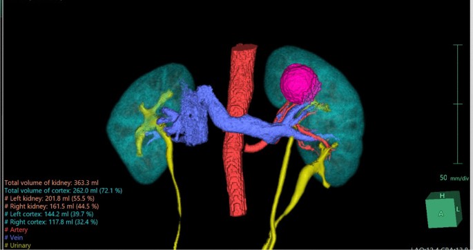
A 45-year-old Vietnamese male patient was incidentally discovered to have a left kidney mass during a routine health check. The patient had a history of hypertension and no other complaints. The CT scan revealed a distinct, confined mass lesion at the upper pole of the left kidney, measuring 32 × 33 × 35 mm, suggestive of RCC. The left kidney was entirely duplex on imaging. We utilized the Fujifilm’s Synapse® Artificial Intelligence (AI) Platform to reconstruct a 3D image of both kidneys and clearly observed the fully duplicated left kidney along with the tumor at the upper pole. Using Tc-99 m DTPA isotope scan, the estimated split renal function of the left kidney was 52.6%, with a glomerular filtration rate of 40.3 ml/minute.
The diagnosis in this case is cT1aN0M0 hilar renal tumor on the entirely duplicated left kidney, Renal score was 9ah. The patient underwent a partial nephrectomy of the left kidney with the assistance of the da Vinci robotic system. During the surgery, we exposed and observed the anatomical structure of the left duplex kidney as seen in the 3D reconstructed image (Fig. 1). The renal tumor appeared distinctly with well-defined borders and was located at the upper pole of the left kidney. We clamped the left renal artery using a Bulldog clamp and proceeded to perform an enucleation of the renal tumor. The warm ischemia time was 23 minute, estimated blood loss was approximately 50 ml, and the total surgical time was 140 minute.
The patient was discharged after 3 days with no postoperative complications observed. The histopathological results confirmed renal cell carcinoma (RCC), with no tumor cells found at the surgical margins, indicating clear resection (Fig. 2). One-month follow-up, the renal function was preserved without change in the serum creatinine level.
Case 2
A 54-year-old Vietnamese female patient was incidentally found to have a right kidney mass during a routine health examination. The CT scan imaging was strongly suggestive for a solid renal mass, likely renal cell carcinoma (RCC). Preoperative tests showed normal results, and both kidneys demonstrated comparable function. The left kidney had a glomerular filtration rate of 50.9 ml/minute, constituting 50.1% as determined by the Tc-99 m DTPA renal isotope study.
With the assistance of Fujifilm’s Synapse® AI Platform, we obtained detailed images of the tumor anatomy, including the blood vessels supplying the tumor. Additionally, the AI platform helped identify the presence of bilateral duplicated kidneys, a detail that was initially overlooked in the conventional CT scan (Fig. 2).
The patient was diagnosed with RCC, clinically staged as cT1bN0M0, and had a RENAL score of 9x. A partial nephrectomy of the right kidney was performed using the da Vinci robotic system. During the surgery, distinct images of the two collecting systems were observed, clearly depicted in the 3D reconstructed image using AI (Figs. 3 and 4). The tumor was prominently located at the lower pole, showing well-defined borders with the normal parenchyma and no invasion into the underlying collecting systems. Examination of the renal hilum revealed a perfect match between the actual branching pattern of the blood vessels and the image generated by Fujifilm’s Synapse® AI Platform.
We clamped the renal artery using a laparoscopic Bull-dog clamp and proceeded to perform an enucleation of the renal tumor. The warm ischemia time was 28 minute, estimated blood loss was approximately 60 ml, and the total surgical time was 150 minute.
The patient was discharged after 4 days with no postoperative complications observed. The histopathological results confirmed renal cell carcinoma (RCC), with no tumor cells found at the surgical margins (Fig. 5). Furthermore, the serum creatinine level was unchanged one month postoperatively.
Discussion
Duplex kidney is the most common congenital anomaly of the urinary system, with a prevalence of approximately 0.8–1% in the general population, often detected incidentally due to the lack of symptoms [5, 6, 11]. Although RCC is the most common kidney cancer, RCC in duplex kidneys is an exceedingly rare condition [4, 6]. In adults, the first reported case of RCC in a duplicated kidney was in 2014 by Mohan et al., involving a 49-year-old male patient who underwent radical right nephrectomy [4]. The first pediatric RCC in a duplex kidney was also found in a 5-year-old girl with cT3aN1M0 disease [5].
In these two cases, we have successfully integrated the latest advancements in medical technology with artificial intelligence and robotic surgery to proactively plan the surgical approach for each patient. This is the first report in the literature highlighting the application of AI in the diagnosis and treatment of RCC in duplicated kidneys with robotic assistance, aiming to preserve renal function.
In this clinical scenario, with the assistance of SYNAPSE 3D, we accurately identified the presence of tumors on the duplicated kidneys. Furthermore, the 3D simulation images helped in identifying the feeding vessels to the tumors and provided a clear visualization of the spatial relationship between the tumors and adjacent structures [1]. This information was crucial in planning the surgical approach effectively. These minimally invasive methods, in addition to ensuring equivalent oncological safety for localized tumors, offer advantages in terms of reduced invasiveness, decreased blood loss, and shorter hospitalization periods [3].
In the realm of medical imaging and preoperative planning, the use of simulation images is paramount. These images aid surgeons to comprehensively understand complex organ structures and intricate vascular systems within the human body. Traditionally, the creation of simulation images involved manual tracing and extraction from computed tomography (CT) scans. However, this method posed challenges, including operator-dependent variations and inconsistencies arising from differences in scan timing and contrast conditions [12]. Such discrepancies could compromise the accuracy of preoperative planning, a critical aspect of surgeries that are performed frequently in clinical practice [13, 14]. The SYNAPSE 3D, an advanced software solution offered by Fujifilm, excel in analyzing target and non-target areas separately and applying density and shape models to images of the target organ. The result is a significant reduction in operator-dependent discrepancies, leading to highly objective simulation images.
The kidney, often likened to a mass of intricate vessels, poses a unique set of challenges for preoperative simulation. Achieving high-quality, accurate simulation images for renal procedures is of paramount importance. These images serve as invaluable guides for surgeons, allowing them to navigate the complex vascular and parenchymal structures of the kidney with confidence and precision [15]. SYNAPSE 3D represents a significant leap forward in preoperative simulation for renal surgeries, particularly robot-assisted partial nephrectomy. This innovative platform streamlines the process of creating 3D images, significantly reducing the time required for constructing detailed vascular maps and predictive states post-tumor excision. The automation of these critical steps empowers surgeons by freeing them to manipulate and verify images during surgery itself [12]. These images derived by 3-D reconstruction may also be helpful for creating models in training simulations for educational purposes [16].
Conclusion
Renal cell carcinoma in congenital duplicated kidneys is a rare condition, and a comprehensive evaluation of renal anatomy is essential for treatment planning. The integration of stimulation imaging AI platform into preoperative planning for robot-assisted partial nephrectomy represents a promising development. These advancements hold promise in complex clinical scenarios to enhance diagnostic precision. Further case series and cohort studies are essential to evaluate the practical applications and the cost–benefit ratio of artificial intelligence-driven 3D reconstruction platforms for standard imaging protocols in future practice.
Data availability
Data supporting the findings of this study are available from the corresponding author upon reasonable request.
References
Lin W-C, Chang C-H, Chang Y-H, Lin C-H. Three-dimensional reconstruction of renal vascular tumor anatomy to facilitate accurate preoperative planning of partial nephrectomy. Biomedicine. 2020;10(4):36.
Hsieh JJ, Purdue MP, Signoretti S, et al. Renal cell carcinoma. Nat Rev Dis Primers. 2017;3(1):17009.
Ljungberg B, Albiges L, Abu-Ghanem Y, et al. European association of urology guidelines on renal cell carcinoma: the 2022 update. Eur Urol. 2022;82(4):399–410.
Mohan H, Kundu R, Dalal U. Renal cell carcinoma arising in ipsilateral duplex system. Turk J Urol. 2014;40(3):185.
Alqarni N, Alanazi A, Afaddagh A, Eldahshan S, Alshayie M, Alshammari A. Renal cell carcinoma in a duplex kidney in pediatric. Urology Annals. 2021;13(3):320.
Domakunti R, Dharamshi J, Dhale A. Renal cell carcinoma coexistence with ipsilateral renal duplex system: a case report. The Pan African Medical J. 2022; 42.
Duong NX, Le M-K, Nguyen TT, et al. Aquired cystic disease – associated renal cell carcinoma: a systematic review and meta-analysis. Clin Genitourinary Cancer. 2024;22:102050.
Nguyen TT, Ngo XT, Duong NX, et al. Single-port vs multiport robot-assisted partial nephrectomy: a meta-analysis. J Endourol. 2024;38(3):253–61.
Singh R, Wu W, Wang G, Kalra MK. Artificial intelligence in image reconstruction: the change is here. Physica Med. 2020;79:113–25.
Agha RA, Franchi T, Sohrabi C, et al. The SCARE 2020 guideline: updating consensus surgical CAse REport (SCARE) guidelines. Int J Surg. 2020;84:226–30.
Yener S, Pehlivanoğlu C, Yıldız ZA, Ilce HT, Ilce Z, Ilce H. Duplex kidney anomalies and associated pathologies in children: a single-center retrospective review. Cureus 2022; 14(6).
Yap FY, Varghese BA, Cen SY, et al. Shape and texture-based radiomics signature on CT effectively discriminates benign from malignant renal masses. Eur Radiol. 2021;31:1011–21.
Zhang H, Yin F, Yang L, et al. Computed tomography image under three-dimensional reconstruction algorithm based in diagnosis of renal tumors and retroperitoneal laparoscopic partial nephrectomy. J Healthc Eng. 2021;2021:3066930.
Ngo XT, El-Achkar A, Dobbs RW, et al. Laparoscopic retroperitoneal heminephrectomy for renal cell carcinoma in horseshoe kidney: a case report and review of the literature. J Med Case Reports. 2023;17(1):512.
Piramide F, Duarte D, Amparore D, et al. Systematic review of comparative studies of 3D models for preoperative planning in minimally invasive partial nephrectomy. Kidney Cancer. 2022; (Preprint): 1–15.
Lin C, Gao J, Zheng H, et al. When to introduce three-dimensional visualization technology into surgical residency: a randomized controlled trial. J Med Syst. 2019;43(3):71.
Acknowledgements
We extend our sincere gratitude to Fujifilm Vietnam Co. for generously providing the Synapse 3D software, which was instrumental in facilitating the three-dimensional anatomical reconstruction for our cases.
Funding
No funding has been received for this study.
Author information
Authors and Affiliations
Contributions
Tuan Thanh Nguyen: Project development, manuscript writing. Minh Sam Thai: Manuscript writing and editing. Quy Thuan Chau: Manuscript reviewing and editing. Ryan W. Dobbs: Manuscript reviewing and editing. Ho Yee Tiong: Manuscript editing. Duc Minh Pham: Manuscript editing. Ho Trong Tan Truong: Manuscript editing. Kinh Luan Thai: Manuscript editing. Huynh Dang Khoa Nguyen: Manuscript writing. Thanh Thien Huynh: Manuscript writing. Huu Phuoc Le: Manuscript editing. Xuan Thai Ngo: Project development, manuscript editing.
Corresponding author
Ethics declarations
Ethics approval and consent to participate
Regarding patient consent statement, the distribution of this publication was discussed and agreed upon as part of the preoperative consent.
Consent for publication
Written informed consent was obtained from the patient for publication of this case report and any accompanying images. A copy of the written consent is available for review by the Editor-in-Chief of this journal.
Competing interests
The authors declare that there are no competing interests regarding the publication of this article.
Additional information
Publisher's Note
Springer Nature remains neutral with regard to jurisdictional claims in published maps and institutional affiliations.
Rights and permissions
Open Access This article is licensed under a Creative Commons Attribution 4.0 International License, which permits use, sharing, adaptation, distribution and reproduction in any medium or format, as long as you give appropriate credit to the original author(s) and the source, provide a link to the Creative Commons licence, and indicate if changes were made. The images or other third party material in this article are included in the article's Creative Commons licence, unless indicated otherwise in a credit line to the material. If material is not included in the article's Creative Commons licence and your intended use is not permitted by statutory regulation or exceeds the permitted use, you will need to obtain permission directly from the copyright holder. To view a copy of this licence, visit http://creativecommons.org/licenses/by/4.0/. The Creative Commons Public Domain Dedication waiver (http://creativecommons.org/publicdomain/zero/1.0/) applies to the data made available in this article, unless otherwise stated in a credit line to the data.
About this article
Cite this article
Nguyen, T.T., Thai, M.S., Chau, Q.T. et al. Three-dimensional reconstruction of renal tumor anatomy for preoperative planning of robotic partial nephrectomy in renal cell carcinoma cases with duplex kidney: a case report. J Med Case Reports 18, 262 (2024). https://doi.org/10.1186/s13256-024-04582-4
Received:
Accepted:
Published:
DOI: https://doi.org/10.1186/s13256-024-04582-4


