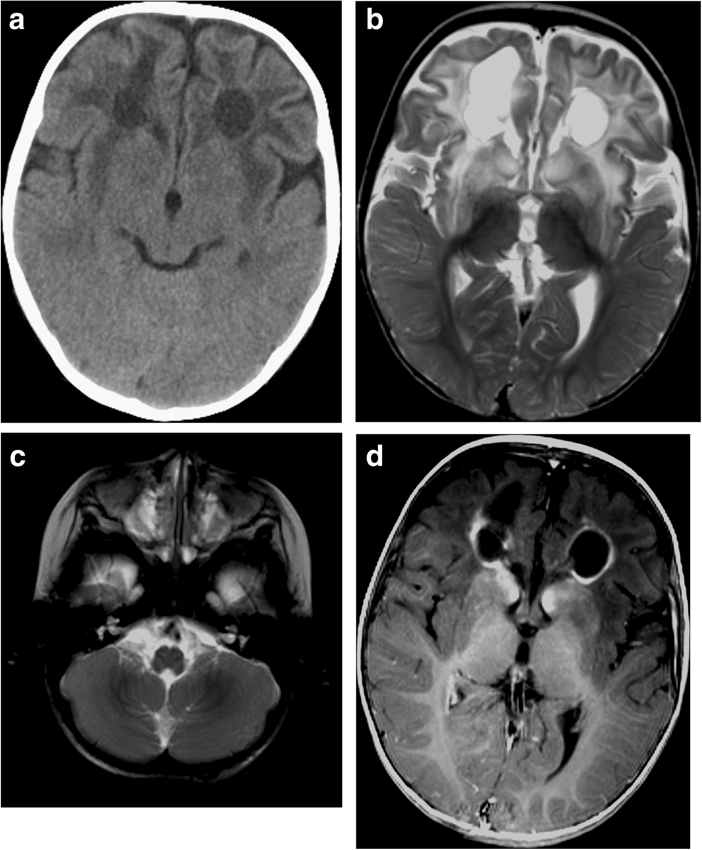
Figure 1

Brain computed tomography and magnetic resonance imaging at 12 months. a Computed tomography image demonstrates low attenuation areas in the white matter of the frontal lobes, putamen, external capsule and claustrum, and cystic formation in the white matter of the frontal lobes. b T2-weighted image shows areas of high signal intensity and cystic formation in the white matter of the frontal lobes, and significant deep gray matter structure abnormality. c T2-weighted image shows small, bilateral hyperintense lesions in the medulla oblongata. d Contrast T1-weighted image demonstrates contrast enhancement in the pericystic lesion and the caudate nucleus.
